Fig. 6.

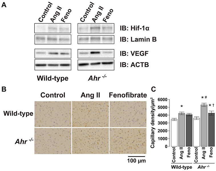
Representative blots of a immunoblot analysis of the amounts of HIF-1α in nuclear extracts from LV tissues and VEGF in protein extracts from homogenates of LV tissues of wild-type and Ahr−/− mice treated with Ang II and fenofibrate. b Capillary detected by immunohistochemical staining with anti-CD31 antibody in the LV wall. Scale bars, 100 μm. c Quantitative analysis of the capillary density in the left ventricle of wild-type and Ahr−/− mice treated with Ang II and fenofibrate. Data are mean ± SEM of six mice per group. *P < 0.05 vs. wild-type control mice; #P < 0.05 vs. wild-type Ang II mice; †P < 0.05 vs. Ahr−/− Ang II mice
