Figure 5.
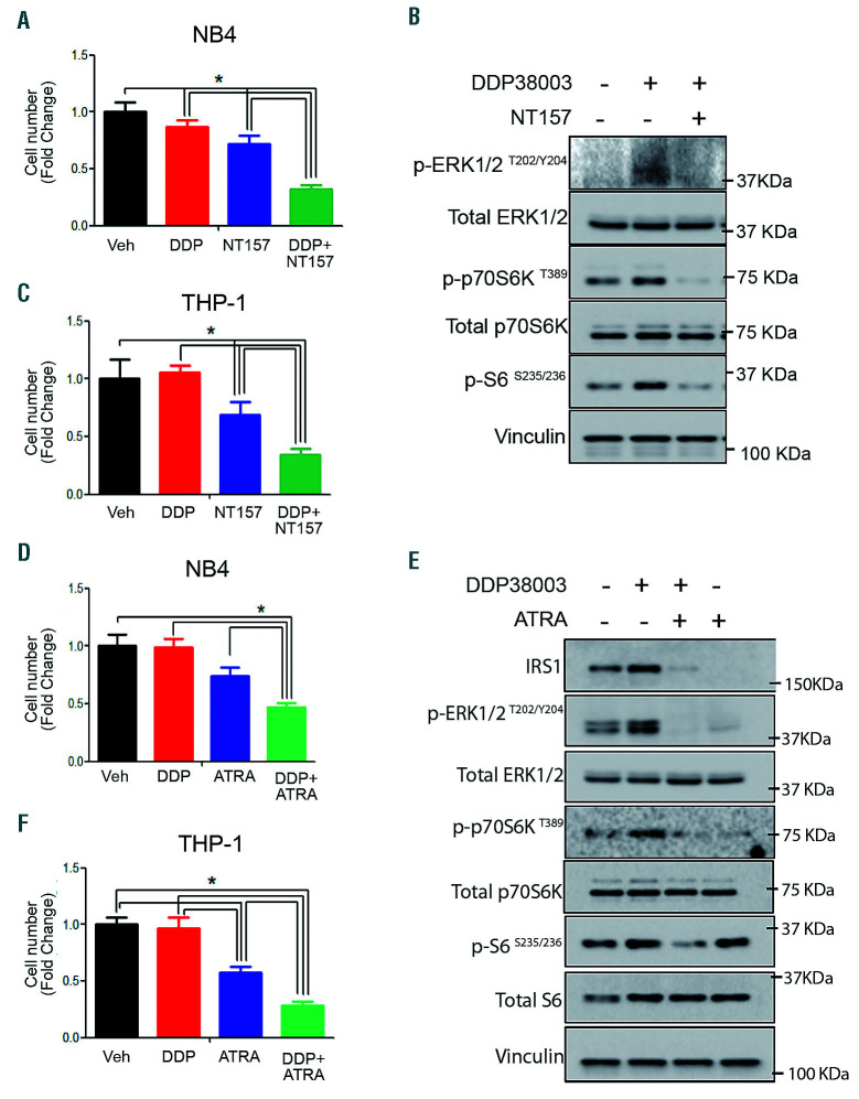
Inhibiting insulin receptor substrate 1 sensitizes resistant acute myeloid leukemia cells to LSD1 inhibition. (A) Relative cell number of NB4 cells treated with either vehicle (Veh), DDP38003 (DDP, 0.5 μM), NT-157 (1.25 μM) or their combination for 72 hours (h). Data were statistically analyzed using one way ANOVA followed by Bonferrroni post hoc test (n=3). *:P<0.05. (C) Relative cell number of THP-1 cells treated with either vehicle (Veh), DDP38003 (DDP, 0.5 μM), NT-157 (1.25 μM) or their combination for 24 h. Data were statistically analyzed using one way ANOVA followed by Bonferrroni post-hoc test (n=3).*: P<0.05. (B) Western blot analysis of THP-1 cells treated as indicated. Vinculin served as a loading control. (D-E) Relative cell number of NB4 (D) and THP-1 (F) cells treated with either vehicle (Veh), DDP38003 (DDP, 0.5 μM), all-trans-retinoic acid (ATRA – 1 μM) or their combination. Data were statistically analyzed using one way ANOVA followed by Bonferrroni post hoc test (n=3).*: P<0.05. (E) Western blot analysis. Vinculin served as a loading control.

