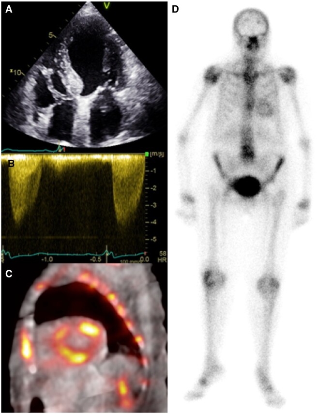
Figure 1.
A 90-year-old man with severe aortic stenosis referred for transcatheter aortic valve implantation. Apical four-chamber echocardiography (A) shows normal biventricular size with mild left ventricular hypertrophy (1.3 cm basal septum). Continuous-wave Doppler confirmed severe aortic stenosis (peak velocity > 4 m/s) (B). The 99mTc-3,3-diphosphono-1,2-propanodicarboxylic acid fused single-photon emission computed tomography/computed tomography short-axis (C) confirms diffuse cardiac retention of tracer, visible also on the planar image (D), compatible with Perugini grade 2.

