Fig. 5.
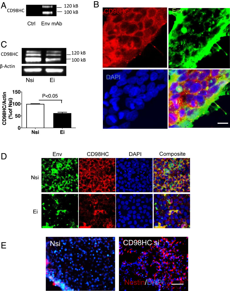
Interaction between HML-2 Env and CD98HC. (A) iPSC lysates were immunoprecipitated using an antibody to HML-2 Env (anti-Env) and analyzed by Western blot using an antibody to CD98HC. Images are representative of three independent experiments (B) Immunostaining of stem cells showed colocalization of CD98HC (red) and HML-2 Env (green). (Scale bar, 10 µm.) (C) Forty-eight hours after transfection with Ei, CD98HC expression was decreased compared with Nsi. Data are presented as mean ± SEM from three independent experiments. (D) iPSCs were transfected with either Nsi or siRNA to HML-2 env. Inhibition of Env with Ei resulted in decreased expression of HML-2 Env (green) and CD98HC (red), as shown by immunostaining. (Scale bar, 20 µm.) (E) iPSCs were treated with either Nsi or siRNA to CD98HC (CD98HC si). Treatment with CD98HC si resulted in an increase in nestin+ cells (red) during neural induction as shown by immunostaining. (Scale bar, 100 µm.) (B, D, and E) DAPI (blue) was used to stain the nuclei. Images are representative of three independent experiments.

