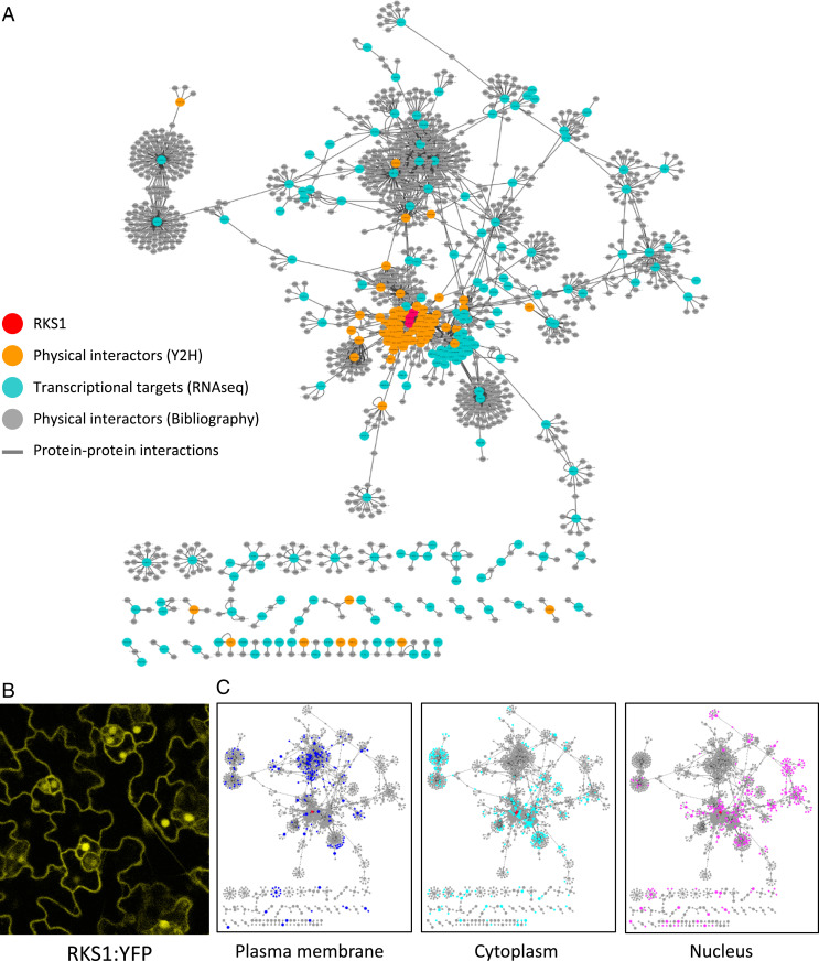
Fig. 3.
Protein–protein interaction network reconstitution reveals a highly interconnected and distributed RKS1-dependent network. (A) The RKS1 protein–protein interaction network plotted with Cytoscape showing the components used for its construction: RKS1 (in red), the physical interactors of RKS1 identified by yeast two-hybrid screening (in orange), the proteins corresponding to the 268 DEGs (in blue), and the proteins identified in the bibliography as experimentally interacting with the proteins corresponding to the 268 DEGs (in gray). (B) Confocal scanning microscopy observation of Arabidopsis leaves of RKS1-OE lines. RKS1 localizes to multiple subcellular compartments: the nucleus, plasma membrane, and cytoplasmic tracks. (C) Display of the subcellular localization of the network components in the compartments where RKS1 has been observed: plasma membrane, cytoplasm, and nucleus.

