Fig. 3.
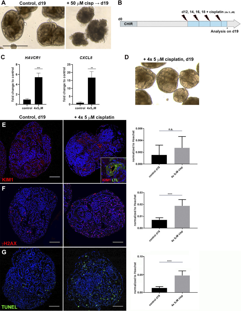
Repeated exposure to low-dose cisplatin reduces cell death and structural deterioration. A: bright-field imaging showing healthy organoids on day 19 (d19) and organoid deterioration after 50 µM cisplatin. B: schematic of repeated low-dose cisplatin treatment. C: quantitative PCR analysis of day 19 organoids showing hepatitis A virus cellular receptor 1 (HAVCR1) and C-X-C motif chemokine ligand 8 (CXCL8) expression increased upon 4 × 5 µM cisplatin. D: organoids treated with 4 × 5 µM cisplatin maintained tubular structures. E–G: immunohistochemistry showing levels of kidney injury molecule-1 [KIM1; colocalized to Lotus tetragonolobus lectin (LTL)+ proximal tubule; E, inset], γH2AX, and TUNEL increased with 4 × 5 µM cisplatin. ns, not significant. **P ≤ 0.01; ***P ≤ 0.001; ****P ≤ 0.0001. Scale bars = 400 μm in A and 100 μm in E–G.

