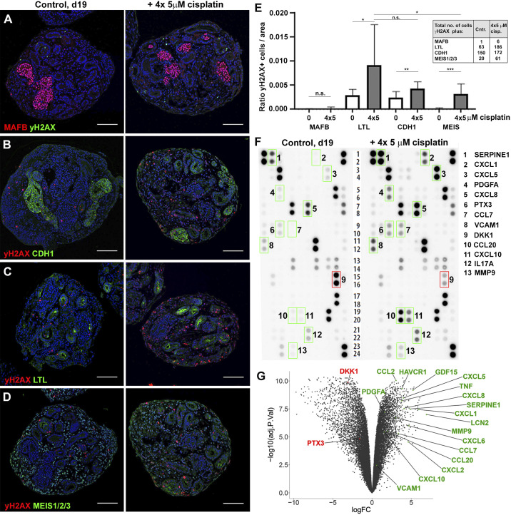
Fig. 4.
Organoids secrete acute kidney injury biomarkers and cytokines in response to cisplatin. A–D: immunohistochemistry on day 19 (d19) control (Cntr) and 4 × 5 µM cisplatin-treated organoids for colocalization of DNA damage (γH2AX) with kidney tissues [MAF BZIP transcription factor B (MAFB) + podocytes, Lotus tetragonolobus lectin (LTL)+ proximal tubules, E-cadherin (CDH1)+ distal tubules, and MEIS1/2/3+ interstitial cells]. E: quantification of γH2AX colocalization with kidney tissues. Inset shows total numbers of double-positive cells counted. F: cytokine array analysis using culture media collected from control and 4 × 5 µM cisplatin-treated organoids. Factors with higher (green boxes) or lower (red box) secretion in cisplatin-treated versus control organoids are shown. G: volcano plot showing the result of RNA sequencing profiling of control and 4 × 5 µM cisplatin-treated organoids. The genes encoding the differentially secreted cytokines and a selection of acute kidney injury biomarkers that are differentially expressed upon cisplatin treatment are highlighted. ns, not significant. See text for abbreviations. *P ≤ 0.05; **P ≤ 0.01; ***P ≤ 0.001. Scale bars = 100 μm.

