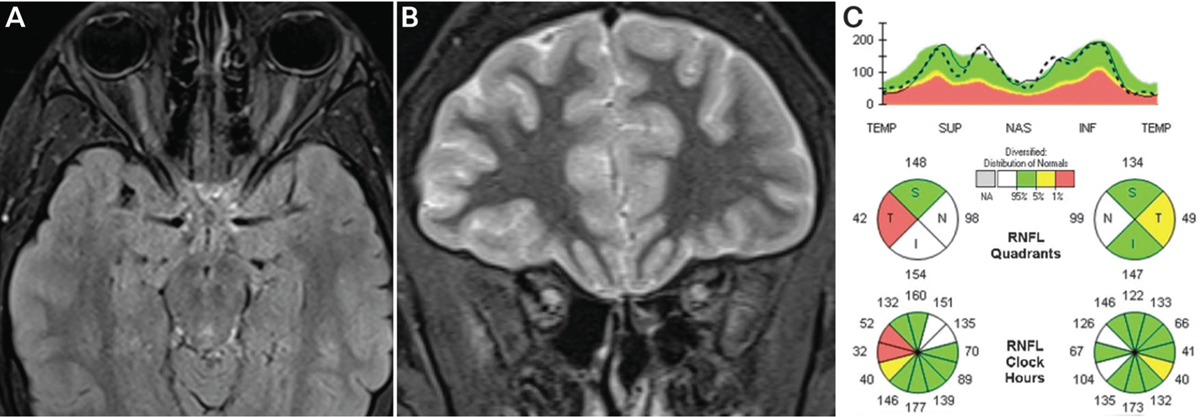
FIGURE 3–4.

Findings of the patient in CASE 3–2. A, Axial fat-saturated T2-weighted turbo inversion recovery magnitude images showing enlarged optic nerves with long T2-hyperintense orbital lesions. B, Coronal short tau inversion recovery (STIR) images showing T2-hyperintense enlarged optic nerves. C, Spectral domain optical coherence tomography of the peripapillary retinal nerve fiber layer showing normal mean thickness in both eyes despite two prior episodes of optic neuritis. Mild segmental retinal nerve fiber layer thickening is seen in the right eye due to edema as well as focal temporal thinning due to prior episodes of optic neuritis, together leading to the normal mean thickness (pseudo-normal mean thickness). The patient had no clinical evidence of disc edema on examination.
