
FIGURE 3–7.
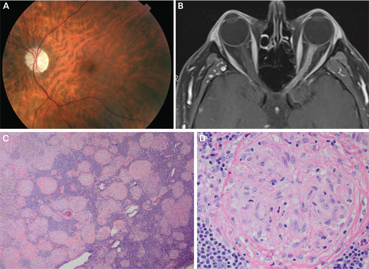
Findings of the patient in CASE 3–3. A, Fundus photography of the left eye revealing optic disc pallor. B, Axial fat-suppressed postcontrast T1-weighted MRI showing longitudinally extensive enhancement of left optic nerve. C, Low-magnification image of hilar lymph node biopsy stained with hematoxylin and eosin (H&E) showing replacement of the normal lymph node architecture by multiple small, well-defined, non-necrotizing granulomas of relatively uniform sizes and shapes that coalesce with variable degrees of fibrosis. D, High-magnification image of granuloma showing cytologic features of the epithelioid histiocytes.
Panels C and D courtesy of Jeffrey Schowinsky, MD.