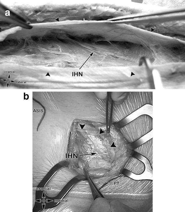
Fig. 6.

Anatomical specimen and a surgical case showing the step 3 landmarks. a After incision of the external oblique aponeurosis (black arrows), the iliohypogastric nerve, accompanied by its nutrient vessel and embedded in its connective tissue, became visible. IHN iliohypogastric nerve, NA nutrient artery. b Surgical case showing the anterior superior iliac spine (ASIS), the pubic tubercle (PT) and the iliohypogastric nerve after incision of the External oblique aponeurosis (black arrow heads)
