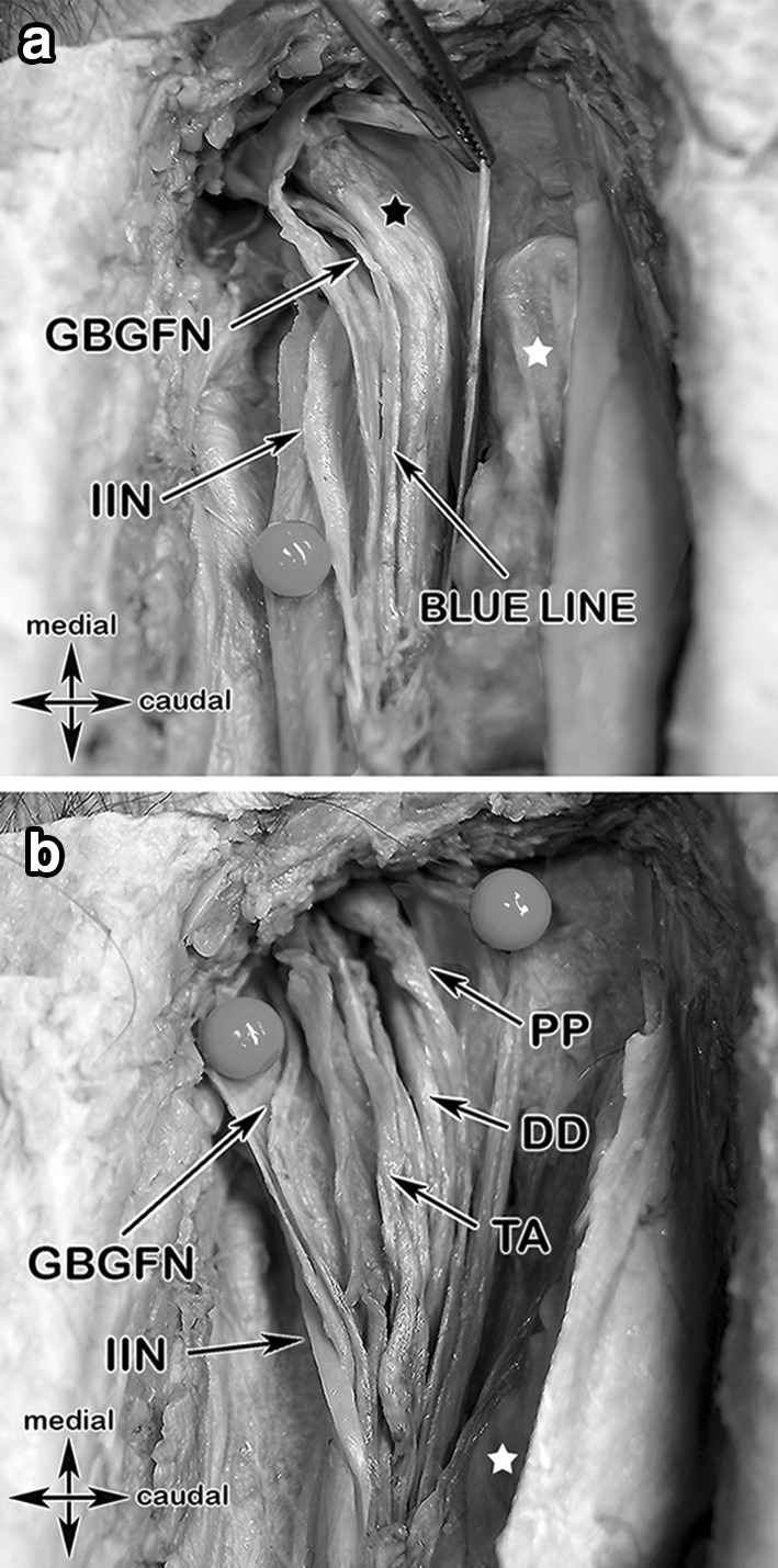
Fig. 8.

Colored anatomical specimen and a surgical case showing the spermatic cord layers and their topographic relationship to important structures in step 4. a The ilioinguinal nerve lying between the external spermatic fascia (purple) and the cremasteric fascia (green), the genital branch of the genitofemoral nerve is travelling along the posterior-medial aspect of the spermatic cord together with the cremasteric vein (“blue line”). b Opening the internal spermatic fascia (pink) exposes the deferens duct, pampiniform plexus and the testicular artery. c Surgical case with exposure of the ilioinguinal (lying dorsal of the spermatic cord), iliohypogastric and the genital branch of the genitofemoral nerves, each marked by a yellow vessel loop
