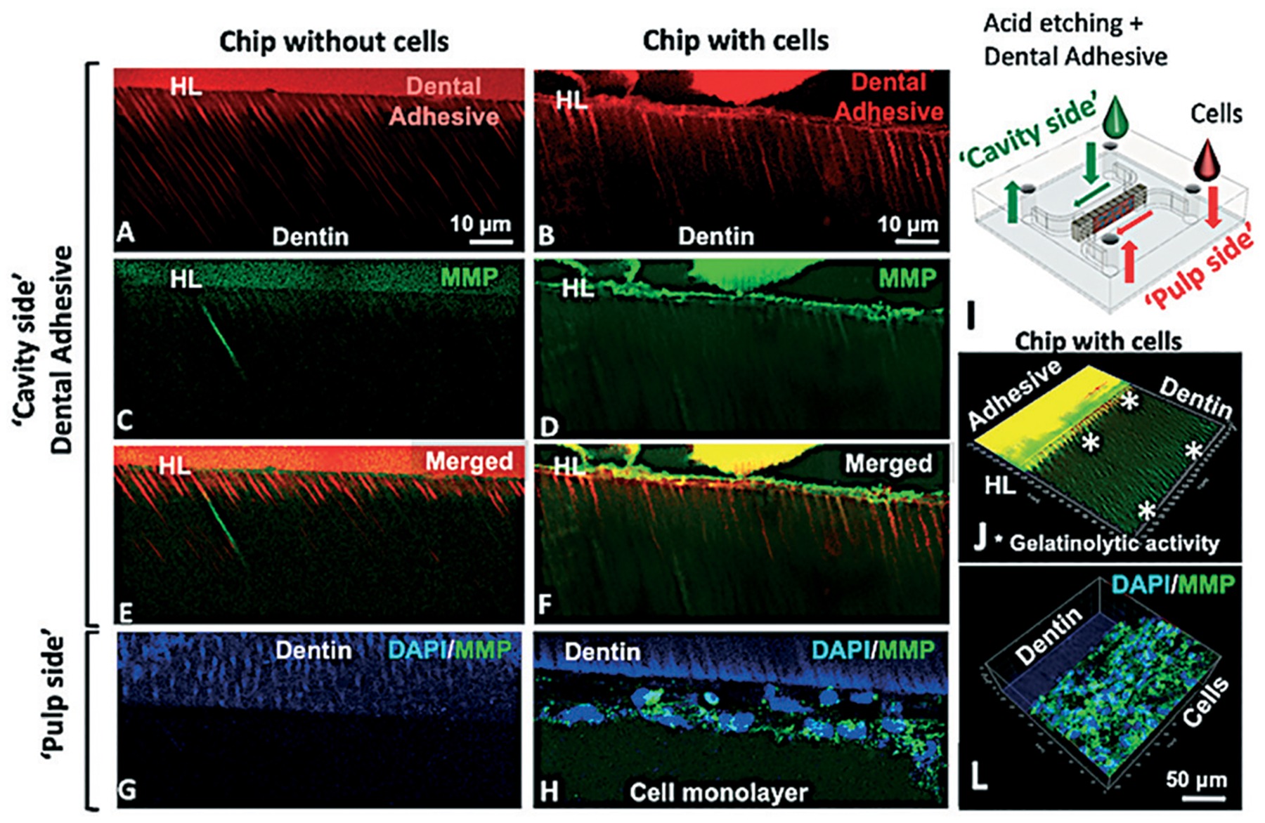
Fig. 7.

Gelatinolytic activity in the hybrid layer on-chip with and without cells after 48 h. Hybrid layer and resin tags present in chips without (A) and with cells (B). Fluorescein-conjugated gelatin showing gelatinolytic activity in the hybrid layer and dentin tubules (C–F). No evidence of gelatinolytic activity on the ‘pulp side’ for chips without cells (G) while for chips with cells, gelatinolytic activity was co-localized with cell cytoplasm (H). Schematic of the chip (I) and 3D orthogonal view of the adhesive side of a chip with cells (J) showing gelatinolytic activity in the hybrid layer inside dental tubules (*) and on the cell side (L), unquenched gelatin co-localized with cell cytoplasm.
