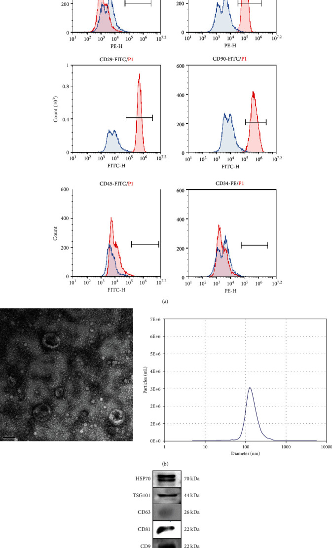
Figure 1.

Characterization of PMSC and PMSC EVs. (a) PMSC phenotypes were tested by flow cytometry. PMSCs were trypsinized and stained with MSC-related markers CD29, CD90, and CD105 and hematopoietic markers CD45, CD34, and HLA. (b) PMSC-EVs were extracted from cell supernatant. Representative micrograph of purified PMSC-EVs under transmission electron microscopy showing a cup-shaped membrane vesicle 30-100 nm in diameter (30,000×). Purified PMSC EVs particles have a diameter of 100 nm. (c). Expression levels of typical molecular markers of EVs were tested by Western blot including HSP70, TSG101, CD63, CD81, and CD9.
