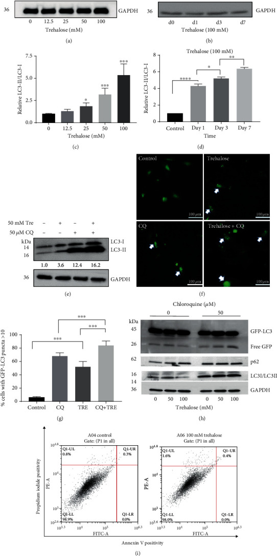
Figure 1.

Trehalose increased autophagosomes formation and autophagy flux. (a–e) Endogenous LC3II expression in cell lysate from ARPE-19 cells treated as designated. (f, g) Live-fluorescence microscopy showing percentage of GFP-LC3–expressing cells containing >10 GFP-LC3 puncta after treatment. (h) Free GFP levels in cell lysate from GFP-LC3–expressing cells after treatment. (i) Flow cytometry results of Annexin V/PI staining in wildtype cells after trehalose treatment. Percentage of late apoptosis and early apoptosis are shown in the upper right and lower right quadrants, respectively. Data represent the mean ± SD of 3 independent experiments. Statistical analysis was performed by one-way ANOVA followed by multiple comparison tests. ∗p < 0.05, ∗∗p < 0.01, ∗∗∗p < 0.001.
