Figure 2.

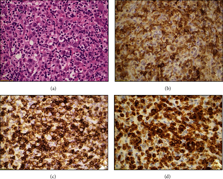
Histologic sections show fragments of soft tissue with extensive infiltration by large, pleomorphic lymphoid cells (a); immunohistochemical studies reveal that neoplastic cells are positive for CD4 (b), CD30 (c), and CD43 (d).

Histologic sections show fragments of soft tissue with extensive infiltration by large, pleomorphic lymphoid cells (a); immunohistochemical studies reveal that neoplastic cells are positive for CD4 (b), CD30 (c), and CD43 (d).