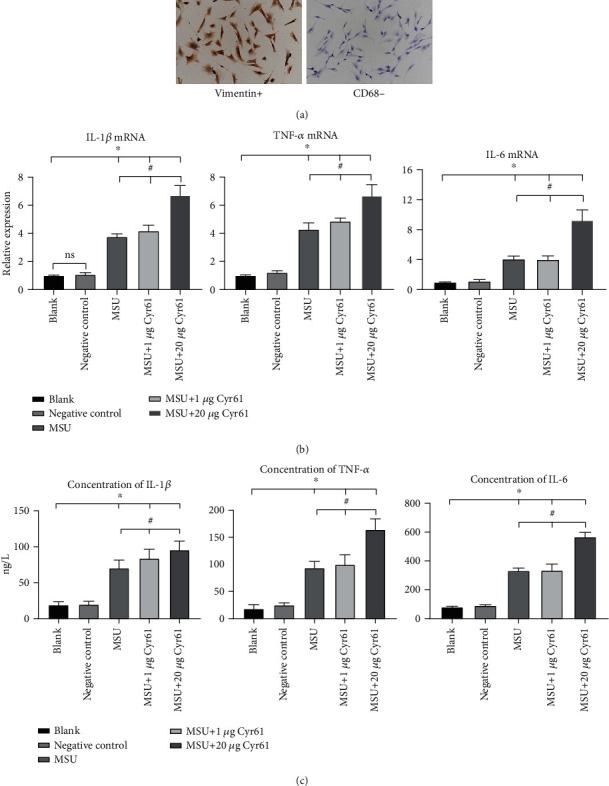
Figure 4.

(a) We used vimentin and CD68 as the rat FLS markers. (b) Relative expression of Cyr61, IL-1β, TNF-α, and IL-6 mRNA in the blank, negative control, MSU-induced, MSU+1 μg Cyr61, and MSU+20 μg Cyr61 groups by PCR. (c) Protein levels of Cyr61, IL-1β, TNF-α, and IL-6 in the same groups by ELISA (∗P < 0.05, vs. blank; #P < 0.05, vs. MSU+20 μg Cyr61).
