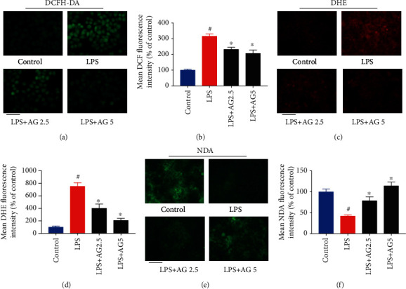
Figure 3.

Andrographolide prevented LPS-induced oxidative stress. (a) Effect of andrographolide on LPS-induced ROS production. The Caco-2 cells were pretreated with andrographolide (2.5 and 5 μM) for 24 h, followed by the treatment of LPS (10 μg/mL) for a further 24 h. After that, the medium was removed and incubated with 10 μM of DCFH-DA for 30 min, and then cells were evaluated by fluorescence microscopy. The scale bar represents 25 μm. (b) The quantitative data of panel (a), and results were expressed as mean DCF fluorescence intensity (n = 6). (c) Effect of andrographolide on LPS-induced superoxide anion radical production. After treatment, cells were collected and incubated with 10 μM of DHE for 30 min, and then cells were evaluated by fluorescence microscopy. The scale bar represents 50 μm. (d) The quantitative data of panel (c), and results were expressed as mean DHE fluorescence intensity (n = 6). (e) Effect of andrographolide on GSH content in LPS-induced Caco-2 cells. After treatment, cells were collected and incubated with 50 μM of NDA for 30 min, and then cells were evaluated by fluorescence microscopy. The scale bar represents 50 μm. (f) The quantitative data of panel (e), and results were expressed as mean NDA fluorescence intensity (n = 6). Data represent means ± SEM (n = 6), #p < 0.05 versus control group, ∗p < 0.05 versus the LPS group.
