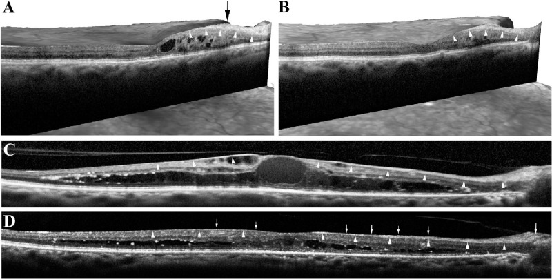
Figure 2.

(A) Three-dimensional OCT scan of an 86-year-old male patient with inferotemporal branch retinal vein occlusion in the right eye. Patient developed macular edema with a peak located 1900 µm nasal to the fovea (black arrow). RNFL is thickened and displays high reflectivity (white arrowheads). (B) Four weeks after intravitreal bevacizumab injection, a horizontal scan from the same meridian reveals resorption of the intraretinal cysts along with a 12.3% decrease in retinal thickness at the peak point (600–525.3 µm). A higher (23.2%; 120–92.1 µm) reduction in RNFL thickness with a decrease in its reflectivity was also observed (white arrowheads). (C) Horizontal line scan through the peak point of the clinically significant DME in the right eye of a 65-year-old female patient. The peak point of the edema was located 5.5 µm nasal and 167.8 µm inferior to the foveola. A diffuse thickening and increased reflectance of the RNFL around the peak point is seen (white arrowheads). (D) Four weeks after an intravitreal aflibercept (2.0 mg/50 mL) injection patient's visual acuity improved from 20/100 to 20/70 along with disappearance of most of the intraretinal cysts. A marked reduction of the RNFL around the peak point was accompanied by loss of diffuse reflectance of the RNFL. At this point, occasional hyperreflective small dots (white arrows) are observed within the RNFL, which may represent aggregates of macromolecules within the axoplasm.
