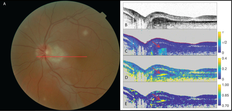
Figure 4.
The left eye of a patient suffering from multifocal choroiditis with doubt among the three retinal specialists (two positive, one negative) as to the presence of subretinal fibrosis. (A) Fundus image; (B) OCT intensity image (colorscale: 0–33 dB); (C) cumulative retardation image (in rad); (D) local birefringence image (in deg/µm); and (E) optic axis uniformity image. Note the elevated birefringent signal in scans (C) and (D) and high optic axis uniformity in (E) in the PS-OCT images (red arrows), indicating the presence of fibrosis.

