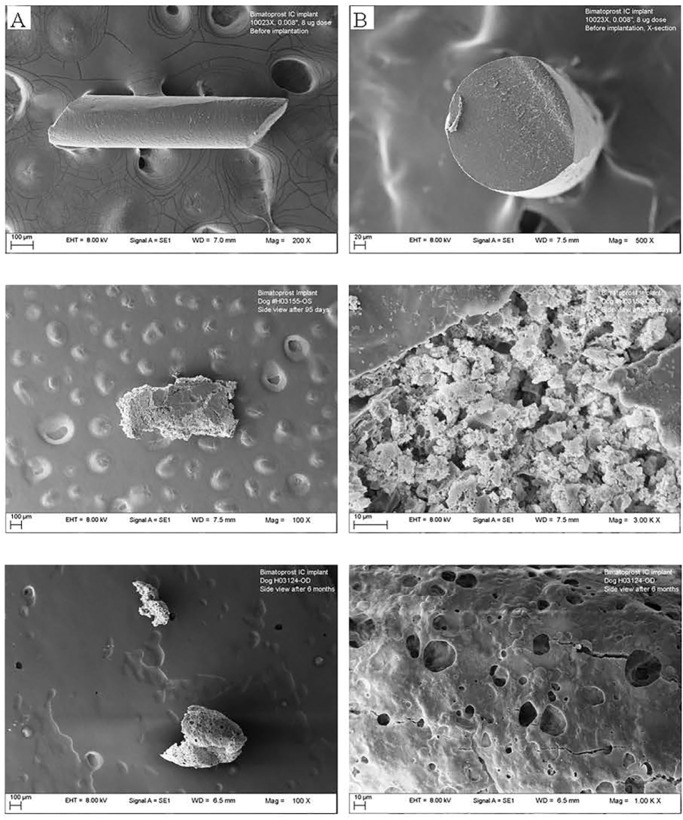
Figure 6.
Representative scanning electron microscopy images of the implant (8-µg dose): (top) before implantation ([A] side view and [B] cross section); (middle) retrieved from the anterior chamber after 3 months (left side view, 100× magnification, right side view, 1000× magnification); (bottom) retrieved from the anterior chamber after 6 months (left side view, 100× magnification, right side view, 1000× magnification).

