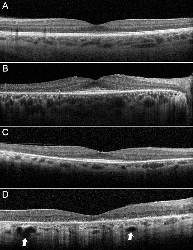
Figure 1.
Choroidal patterns in RP. If compared with a control participant (A), pattern 1 is characterized by normal-appearing choroidal vessels (B). Pattern 2 disclosed reduced Haller and Sattler layers (C). Pattern 3 showed also the presence of choroidal caverns (white arrows) (D). It is worth noticing the increase of choroidal hyperreflective signal, interpretable as an enlargement of a stromal component.

