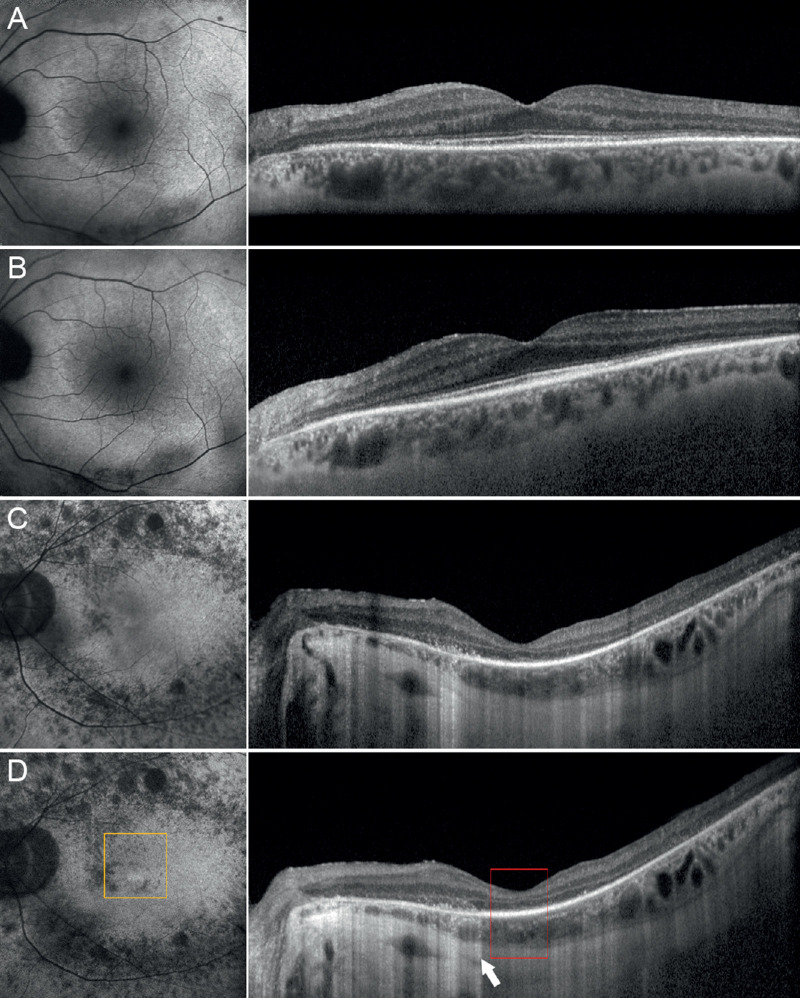
Figure 3.
One-year follow-up and RP progression. Pattern 1 showed unremarkable changes if looking at baseline features (A) and 1-year follow-up (B). On the contrary, pattern 3 showed a remarkable progression of outer retinal alterations, if comparing baseline (C) with 1-year follow-up (D), with increased window effect on structural OCT (white arrow), EZ disruption (red square), and fundus autofluorescence alterations (orange square).

