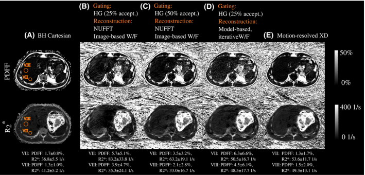
Figure 3.

PDFF (top) and (bottom) maps of a 25‐year old patient (male, BMI: ) from (A) the breath‐held Cartesianeference scan, and (B)‐(E) the free‐breathing stack‐of‐stars acquisition (end‐expiratory frame). The radial dataset was reconstructed (B) using motion‐gating (25% acceptance rate) followed by NUFFT and image‐based water‐fat separation, (C) using motion‐gating (50% acceptance rate) followed by NUFFT and image‐based water‐fat separation, (D) using motion‐gating (25% acceptance rate) followed by model‐based water‐fat separation ("Motion‐resolved XD" with ), and (D) using motion‐resolved XD reconstruction. Two exemplary VOIs were drawn in the parameter maps of “BH Cartesian" in the Couinaud liver segments VII and VIII. HG, hard‐gating
