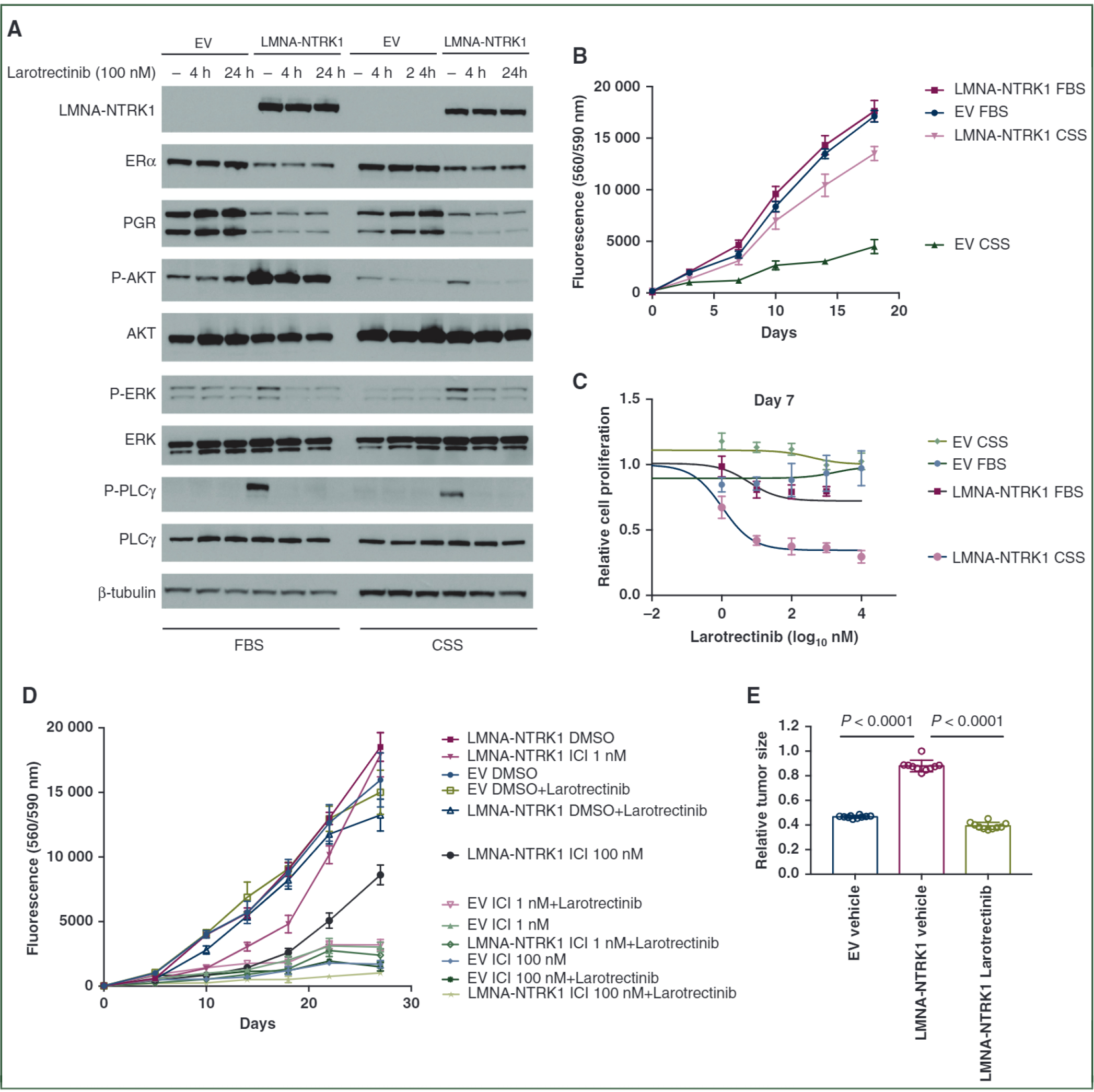
Figure 2. LMNA-NTRK1 fusion promotes hormone-independent growth and confers resistance to endocrine therapy.

(A) Western blot analysis of ERα, PgR and phosphorylation of AKT, ERK and PLCγ in MCF7 cells expressing LMNA-NTRK1 fusion that were cultured with either 10% FBS or 10% CSS and treated with 100 nM larotrectinib for 4 h or 24 h. (B) LMNA-NTRK1 fusion promoted hormone-independent growth of MCF7 cells. MCF7 cells with empty vector (EV) or LMNA-NTRK1 fusion were seeded into 96-well plates in DMEM/F12 medium supplemented with either 10% FBS or 10% CSS, and cell proliferation was assayed with resazurin. (C) Proliferative inhibition of MCF7 cells expressing LMNA-NTRK1 fusion by larotrectinib. MCF7 cells with EV or LMNA-NTRK1 fusion were treated with various doses of larotrectinib in medium supplemented with 10% FBS or 10% CSS. (D) LMNA-NTRK1 fusion confers resistance to fulvestrant. MCF7 cells with EV or LMNA-NTRK1 fusion seeded into 96-well plates were treated with various doses of fulvestrant (ICI) and/or 100 nM larotrectinib. (E) LMNA-NTRKl-expressing MCF7 xenografts after estrogen deprivation. MCF7 xenograft tumors expressing LMNA-NTRK1 fusion were established in nude mice. Then the estradiol pellets were removed, and the mice were treated with either vehicle or 200 mg/kg of larotrectinib via gavage once daily for 2 weeks. The mice bearing MCF7 tumors expressing the EV were treated with vehicle as control. The relative tumor volume compared with the volume measured before treatment of each group ± standard deviation (n = 10 mice/group) is shown with t-test P values.
CSS, charcoal-stripped bovine serum; DMEM, Dulbecco’s Modified Eagle Medium; DMSO, dimethyl sulfoxide; ERα, estrogen receptor alpha; FBS, fetal bovine serum; PgR, progesterone receptor.
