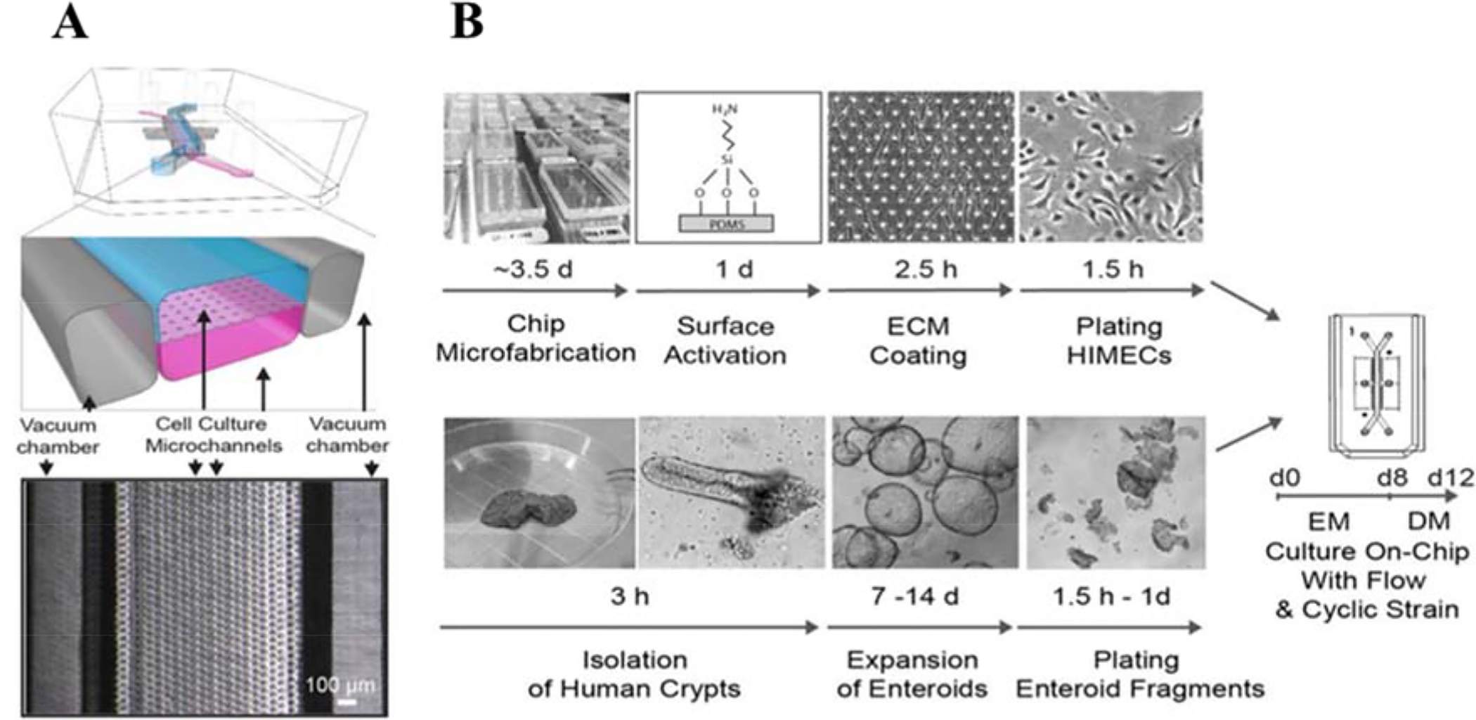
Figure 5.

Primary human Intestine Chip platform. (A) A schematic illustration of the chip and a top view phase contrast micrograph of the chip. Vacuum chambers were incorporated to facilitate peristalsis-like mechanical deformation of the tissue, which has been shown to aid in formation and maturation of 3D tissue structures. (B) Schematic illustration of the step-by-step procedure for the establishment of on-chip co-cultures of primary human intestinal epithelium and intestinal microvascular endothelium. Reproduced from [88], with permission from Nature Publishing Group.
