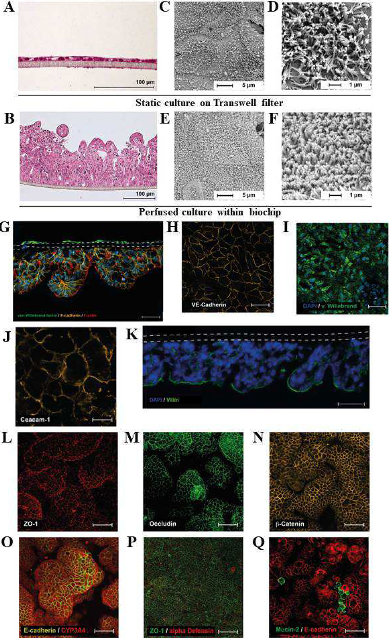
Figure 6.

Mechanically stimulated cells form crypt and villus-like structures and express endothelial and intestinal epithelial markers. A, B) Histological H&E staining of Caco-2 cell layers cultured A) statically in the Transwell, and B) under perfusion in the biochip. C-F) Scanning electron microscopy of Caco-2 cell layers cultured for 7 days under C, D) static conditions on Transwell filters, and E, F) under perfusion in the biochip. G) Cross-section of the three-dimensional intestinal model perfused at 50μl/min (in both channels): endothelial cells express von Willebrand factor (green), epithelial cells express E-cadherin (orange), and F-actin (red). Both cell layers are separated by a porous membrane (dashed line). Actin filaments are stained with phalloidin (red). Scale bar 100 μm. Nuclei were stained with DAPI (blue). H-I) Endothelial cells form a confluent monolayer and express H) VE-cadherin (orange) and I) von Willebrand factor (green). J-Q) Epithelial cell layer: Expression of J) CEACAM-1 (orange); K) villin (green) (DAP blue, dashed lines marks membrane); L) ZO-1 (red); M) occludin (green); N) β-catenin (orange); O) E-cadherin (orange); CYP3A4 (red); P) α-defensin (red); ZO-1 (green); Q) mucin-2 (green); E-cadherin (red); Nuclei were stained with DAPI (blue). Figure reproduced from Ref. [105], with permission from Elsevier.
