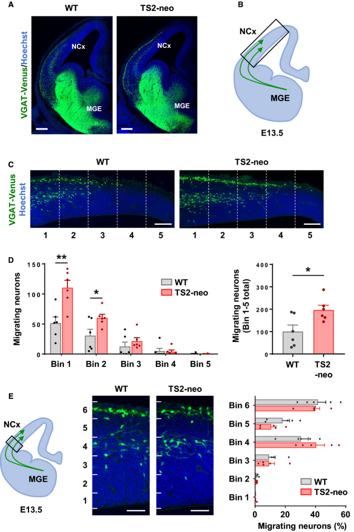
Fig. 4.

Excess migrating inhibitory neurons in TS2‐neo embryonic brain. (A) Coronal sections of the telencephalon at E13.5. Immature inhibitory neurons were labeled with Venus. (B) Schematic illustration of tangential migratory streams derived from the medial ganglionic eminence (MGE) to the neocortex (NCx). The box shows the region of interest presented and analyzed in C and D. (C) Representative images of migrating neurons in the embryonic neocortex. (D) Equidistance bin analysis revealed an increase in migrating neurons labeled with Venus that reached the neocortex in TS2‐neo mice (n = 6 embryos per group). (E) Number of Venus‐positive neurons in radially aligned six equidistance bins (n = 6 embryos per group). Data are mean ± SEM, *P < 0.05, **P < 0.01; unpaired two‐tailed t‐test with Welch's correction (B, C). Scale bars, 200 μm (A), 100 μm (C, E).
