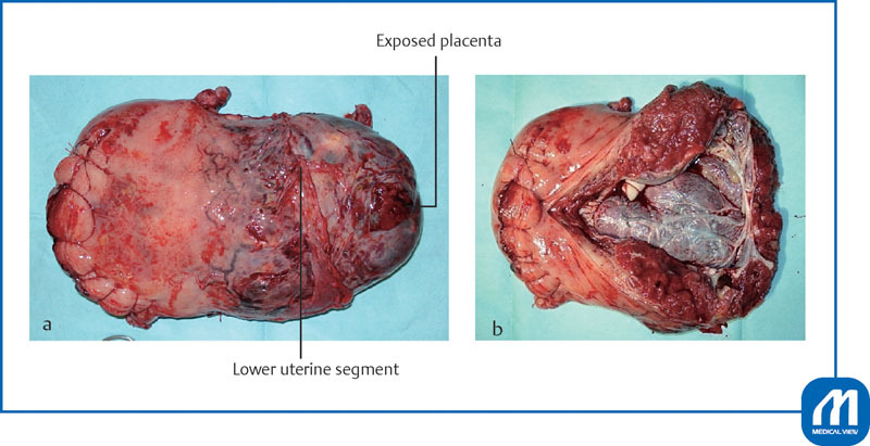
Fig. 11.

Surgical specimen of the uterus with placenta percreta. The fetus was delivered by transverse incision in the uterine fundus. The lower part of the uterus was enlarged, showing a potbelly-like protrusion, in a broad range due to placental attachment. The ureter was located extremely close to the uterus. From the time of separation of the bladder, blood flow was blocked twice for 30 minutes with a balloon occlusion catheter to perform total hysterectomy. The placenta was exposed in the area of bladder separation, leading to a pathological diagnosis of placenta percreta. The amount of intraoperative bleeding was 1,500 mL. Autologous blood alone, a quantity of 500 mL, was transfused, and the surgery was completed. ( a ) Anterior uterine view; ( b ) Posterior uterine view. The posterior uterine walls were cut to open the uterine cavity. See the placenta to invade into the anterior uterine lower segment around the cesarean section scar. (Reproduced with permission from Takeda S. Cesarean section for placenta previa and placenta previa accrete spectrum. In: Hiramatsu Y, Konishi I, Sakuragi N, Takeda S, eds. Mastering the Essential Surgical Procedures OGS NOW, No.3. Cesarean section. (Japanese). Tokyo: Medical View; 2010:102–115. Copyright © Medical View).
