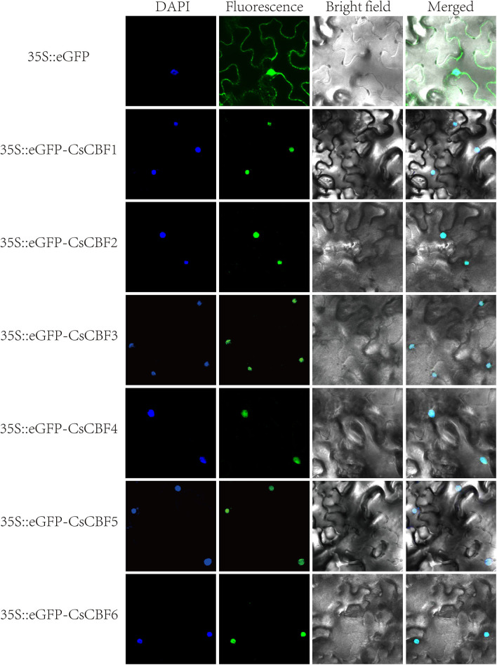
Figure 4.
Subcellular localization of CsCBF proteins in tobacco epidermal cells. The 35S::GFP vector was used as a positive control. Images show 4',6-diamino-phenylindole (DAPI) staining fluorescence, GFP fluorescence, and bright light individually and in combination to demonstrate the morphology of the cells.

