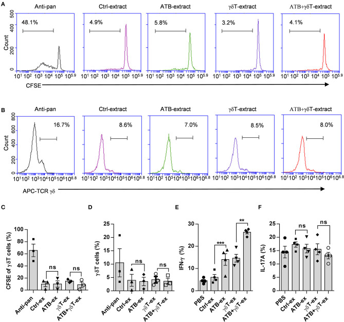
Figure 4.
Cecum contents from ATB-treated mice do not contribute to the proliferation of γδT cells. (A) Representative plot of γδT cell proliferation. Peripheral blood mononuclear cells (PBMCs) isolated from healthy donors were stained with carboxyfluorescein succinimidyl ester (CFSE) and cultured in wells coated with anti-pan γδTCR or treated with fecal dilutions from different mice. (B) Representative plot of γδT cell frequencies in wells under the same conditions described in (A). (C) Statistical diagram based on three experiments of γδT cell proliferation in (A), ns, no significant. (D) Statistical charts of γδT cell purity based on three experiments of (B). ns, no significant. (E) Interferon gamma (IFN-γ) secreted in the supernatant of γδT cells was detected using the same methods as described in Figure 3D. Student's t-test. **p < 0.01, ***p < 0.001. (F) Interleukin (IL)-17A secreted in the supernatant of γδT cells was detected using the same methods as described in Figure 3D, and the data represent three independent experiments, ns, no significant (ANOVA).

