Figure 3.
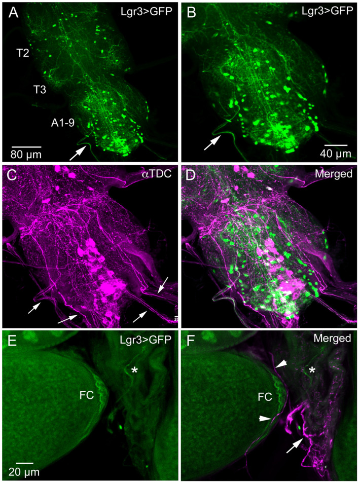
Lgr3-Gal4 and octopamine expressing efferent neurons innervate oviduct muscle. (A) Overview of adult thoracic (T2, T3) and abdominal (A1-9) neuromeres with Lgr3-Gal4 expressing neurons. One efferent axon emerging from the abdominal neuromeres is seen at arrow (Lgr3VP16-Gal4). (B–D) The same specimen, which has also been immunolabeled with antiserum to Tdc2 (tyrosine decarboxylase; magenta). Note that the peripheral Lgr3-expressing axon at large arrows does not colocalize Tdc2 and Lgr3 labeling. Also note that quite a few Tdc2 immunolabeled peripheral axons emerge from the ventral nerve cord (small arrows), some probably destined for the oviduct muscle. (E,F) Part of the ovary and oviduct muscle with Lgr3 (Lgr3VP16-Gal4) and Tdc2 expression. Basal follicle cells (FC) express Lgr3-Gal4 and so does one axon on oviduct muscle (at asterisk). In (F) several axons immunolabeled with anti-Tdc2 are seen on oviduct muscle (arrow and arrow heads). No colocalization of Lgr3 and Tdc2 could be detected.

