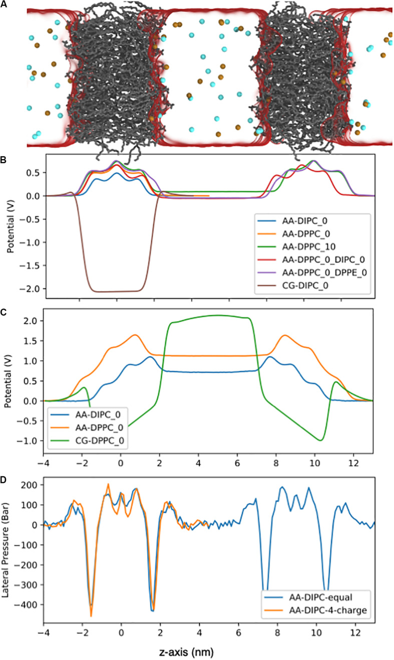
FIGURE 9.
Electrostatic potential for the AA lipid membranes. (A) Snapshot of the double bilayer set-up. The DPPC lipids are shown as gray lines, ions are red and blue balls, and water is a translucent surface. (B) Bilayers with no charge imbalance between aqueous compartments. The potentials were set to zero on the left side of the bilayer. (C) Double bilayer systems with a 4e charge imbalance. Water regions have a flat potential due to conducting counter ions. (D) Lateral pressure profile for the DIPC double bilayer with and without a 4e charge imbalance.

