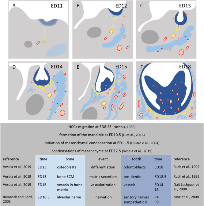
SCHEME 2.

Development of mesenchymal condensations in mouse mandible. Figures show the mandible at stage ED11 (A), ED12 (B), ED13 (C), ED14 (D), ED15 (E), ED18 (F). Dental condensation (dark blue), bone condensation (light blue), Meckel’s cartilage (dark gray), nerves (yellow), vessels (red).
