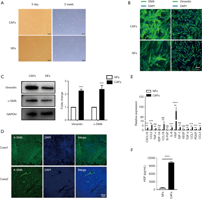
Figure 1.
HGF was up-regulated by CAFs in HCC. (A) Representative images showed the fibroblasts morphology in vitro. The two pictures on the left show freshly isolated fibroblasts from the tissues, while the two images on the right show fibroblasts cultured in vitro for 2 weeks; (B,C) immunofluorescence staining and western blot showing the expression of α-SMA and vimentin in NFs and CAFs; (D) representative immunofluorescence images showing two HCC cases with high α-SMA expression (case 1) and low α-SMA expression (case 2); (E) qRT-PCR indicated mRNA expression differences of soluble factors that CAFs and NFs secreted; (F) CM from CAFs and NFs was collected, and the concentration of HGF was determined using human HGF ELISA. CAFs secreted a significant amount of HGF (9,000, 12,000 pg/mL). Data are presented as the means ± SEM of three independent experiments, the quantitative analysis are done for western blot. ns: not significantly different. **, P<0.01; ***, P<0.001; ****, P<0.0001, t-test. HGF, hepatocyte growth factor; CAF, cancer-associated fibroblast; HCC, hepatocellular carcinoma; α-SMA, α-smooth-muscle actin; NF, normal fibroblast; qRT-PCR, quantitative real-time polymerase chain reaction; CM, conditioned medium; ELISA, enzyme-linked immunosorbent assay.

