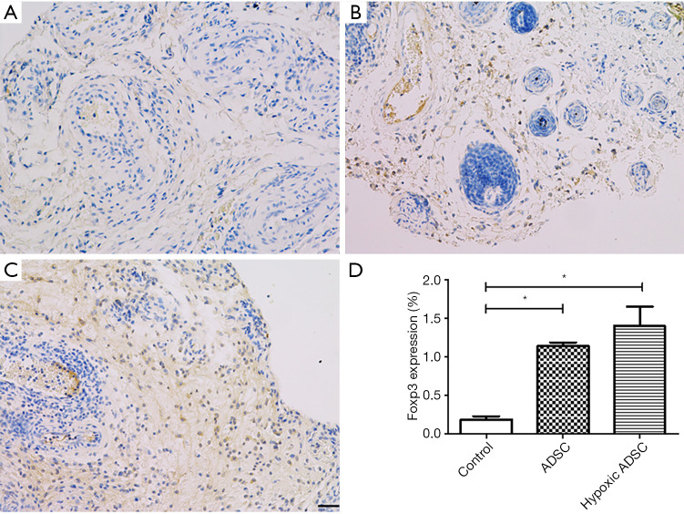
Figure 5.
Recipient ADSCs induced regulatory T-cell expression in allografts. Representative images of immunohistochemical staining of the allograft skin tissue in the control group (A), ADSC treated group (B) and hypoxia primed ADSC treated group (C). (D) A significant increase in Foxp3 expression in the subcutaneous layers of the ADSC and hypoxic ADSC treated groups was observed, compared with the media control on postoperative day 14. (*, P<0.05) (Scale bar =50 µm). ADSC, adipose-derived stem cell; Foxp3, Forkhead box p3.

