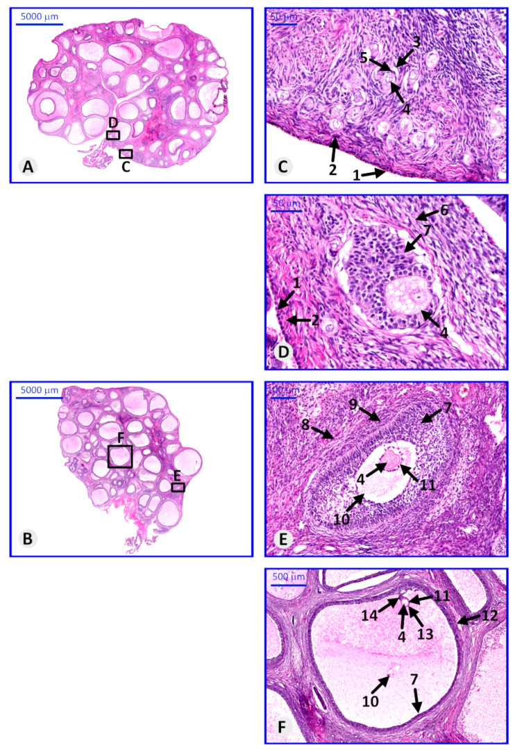
Figure 3.
Paraffin section of porcine ovary stained with hematoxylin and eosin (H&E), representing the histological structure of ovary and follicles in particular stages of maturation. (A,B)—representative ovaries with visible follicles in small magnification; (C)—primordial follicles, (D)—primary follicle, (E)—secondary follicle, (F)—Graafian follicle. Arrows: 1—germinal epithelium, 2—tunica albuginea, 3—primordial follicle, 4—oocyte, 5—follicular cells, 6—primary oocyte, 7—granulosa cells, 8—secondary follicle, 9—tunica interna and externa, 10—antrum, 11—zona pellucida, 12—Graafian (mature) follicle, 13—corona radiate, 14—cumulus oophorum.

