Figure 4.
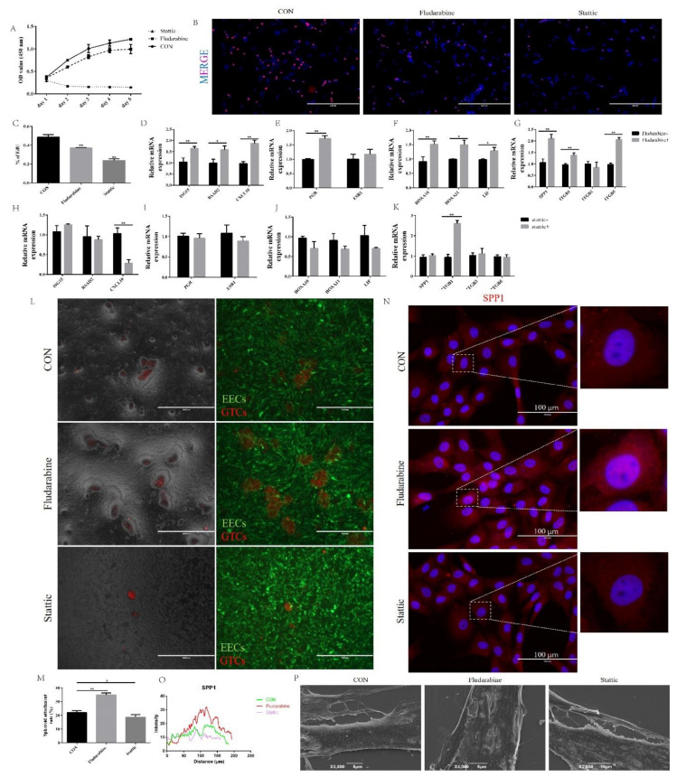
Inhibition of the STAT1 pathway suppressed BCL2L15 silencing-induced endometrial receptivity defects. (A) The cell proliferation was measured using a CCK-8 assay. (B,C) The DNA synthesis was measured using an EdU proliferation assay (D–K) The shBCL2L15 EECs were pretreated with or without fludarabine and Stattic. Then, the EECs were treated with hormones, followed by treatment with IFN-τ for 12 h, and then collected for real-time quantitative PCR. (L,M) Fludarabine treatment increased the adhesion of GTCs spheroids on EECs. Scale bar = 1000 μm. (N,O) Fluorescence microscope images of the SPP1 expression in the CON, fludarabine, and Stattic EECs, which were treated with hormones followed by IFN-τ treatment for 6 h. Representative images of three independent experiments are shown. Scale bar = 100 μm. (P) Scanning electron microscopy (SEM) of EECs, which were pretreated with or without fludarabine and Stattic. Then, EECs were treated with hormones followed by treatment with IFN-τ for 12 h. The data are presented as the means ± S.E.M. of three independent experiments. * Significant difference (p < 0.05) compared with other groups; ** Significant difference (p < 0.01) compared with other groups.

