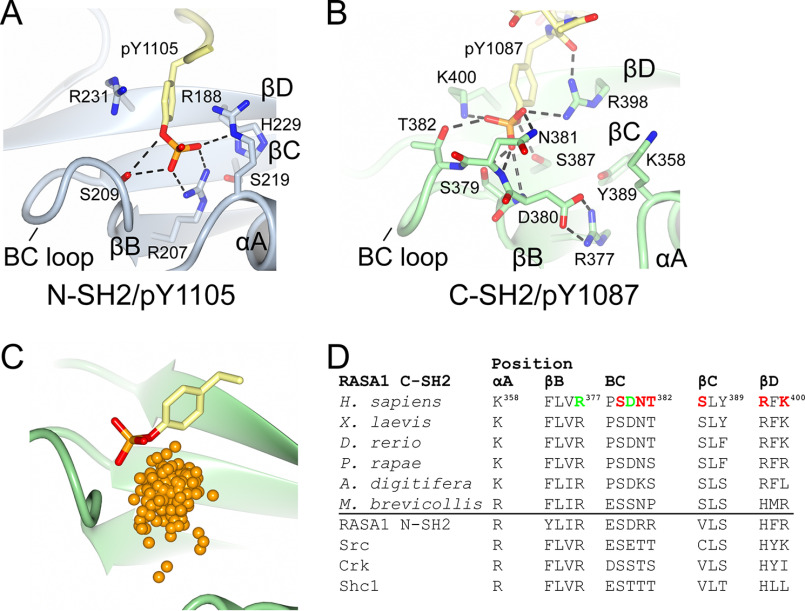
Figure 3.
Phosphotyrosine-binding sites of p120RasGAP. A and B, detailed binding site interactions of the phosphotyrosine to p120RasGAP N-SH2 (PDB code 2PXC) (39) (A) and p120RasGAP C-SH2 (B). C, comparison of phosphotyrosine bound to p120RasGAP C-SH2 with the location of the phosphate atom in 245 phosphotyrosine-bound SH2 domains identified by the Dali server. Locations of the phosphates are shown as orange spheres. SH2 domains identified and superposed using by the Dali server (246 contain phosphotyrosine; 2BBU not included in analysis). D, conservation of key binding site residues over evolution and in human SH2 domains. pTyr-binding residues are colored red, and the FLVR arginine–Asp380 salt bridges are colored green.

