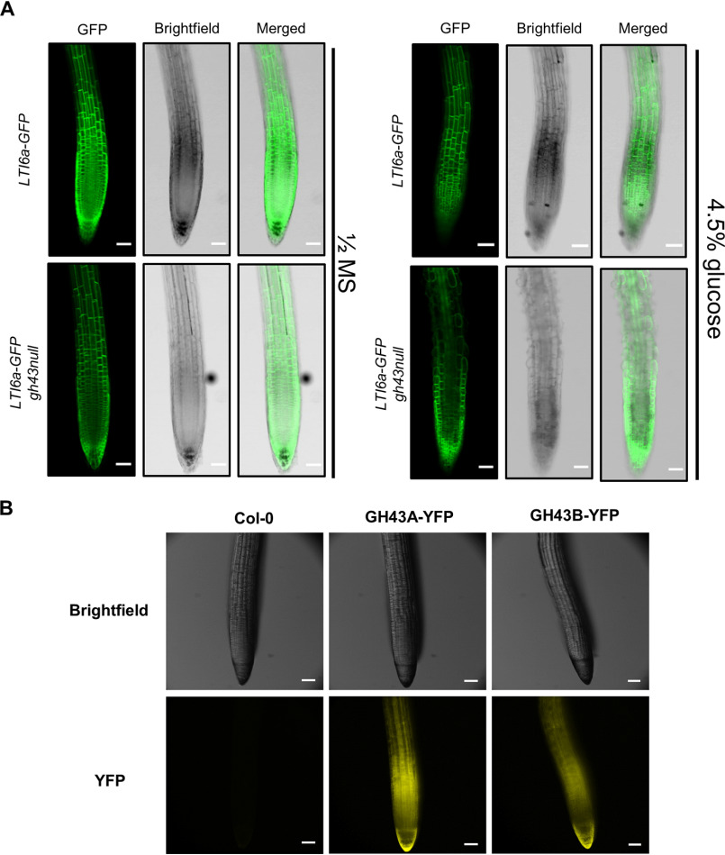
Figure 2.
The initiation of the sugar-induced root swelling in gh43null. A, confocal laser scanning microscope images of Col-0 and gh43null roots stably expressing the LTI6a-GFP plasma membrane marker. Images were obtained 10 h after moving 4-day–old seedlings to nutrient media without (left) and with 4.5% glucose (right). Scale bars = 100 μm. B, fluorescent stereomicroscope images of native promoter GH43-YFP signal in roots. Seedlings were grown on nutrient media without sugar for 4 days. Scale bars = 100 μm.

