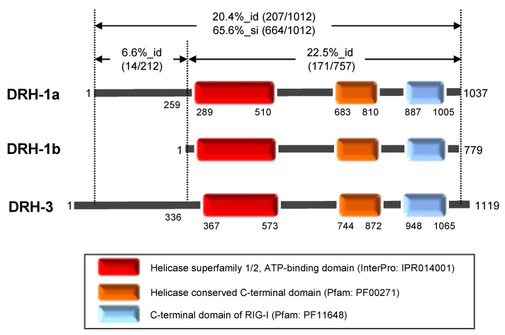
Figure 1.
The Caenorhabditis elegans Dicer-related helicase (DRH) proteins prepared in this study. The schematic structures of DRH-1 isoforms a and b and of DRH-3 are shown. Numbers indicate the amino acid residue positions. DRH-1 isoform b lacks the N-terminal 258-amino acid region that is present in isoform a. The three domains conserved in DRHs and the positions of their amino acid residues are indicated by the box outlines. The percent identity (%_id) and/or similarity (%_si) in the entire region as well as the N-terminal and the central-to-C-terminal regions between DRH-1a and DRH-3 are indicated as the number of identical or similar residues in the total number of residues in the region.

