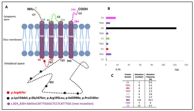
Figure 1.
Location of identified mutations on the Peripherin-2 (PRPH2) protein and the frequency and location of all reported PRPH2 gene mutations. The PRPH2 protein scheme shows the location of the identified mutations. (A) The PRPH2 peptide chain shows extradiscal (C1, C2, and C3), transmembrane (TM1, TM2, TM3, and TM4), and intradiscal space locations (ID1 and ID2). The numbering indicates the amino acid positions at the boundaries of the domains. The mutations identified in the patients are indicated by circles (i.e., red, p.Arg46Ter; black, p.Lys154del, p.Gly167Ser, p.Arg195Leu, p.Val209Ile, and p.Pro216Ser; and purple for the novel mutation c.824_828+3delinsCATTTGGGCTCCTCATTTGG). (B) Representation of the location (C) and the frequency and number of all reported PRPH2 mutations are based on the Human Gene Mutation Database (HGMD 2020.1).

