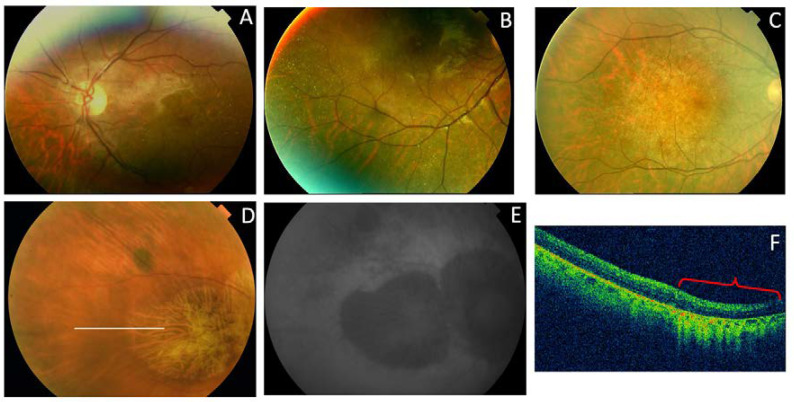
Figure 14.
The clinical appearance of family 5c (p.Arg195Leu). (A) The posterior pole and (B) mid-periphery from F5c-VI-2 had very small whitish dots. (C) F5c-V-2 had a more advanced appearance. (D) F5c-IV-4 had macular atrophy consistent with the diagnosis of CACD, with evident atrophy in the (E) AF and (F) SC-OCT (red brace) images.

