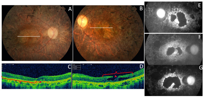
Figure 17.
The RP phenotype in the p.Pro216Ser PRPH2 gene mutation from F7-III-1. (A,B) The ocular fundus had pigmented spiculae in the periphery; the SD-OCT images (C,D) and autofluorescent images show atrophic macular areas of the LE (E) and RE (F) at the age of 48 years that increased 19 years later in the RE (G), the photopic ffERG was unrecordable at this moment.

