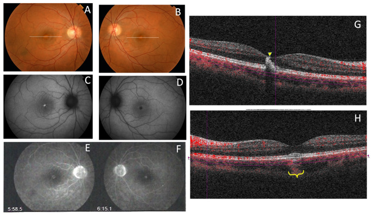
Figure 19.
The clinical appearance of the case from F8-II-2 with the new mutation, c.824_828+3delinsCATTTGGGCTCCTCATTTGG. The fundus appearance of AVMD shows (A,B) yellow foveal deposits, which is (C) hyperautofluorescent in the RE and (D) hypofluorescent in the LE; (E,F) FA shows hyperfluorescence of the lesion in both eyes and (G,H) SD-OCT shows hyper-reflective subfoveal deposits bilaterally that (G) extend to the inner retina of the RE (yellow arrow) and (H) cause choroidal hyper-reflectivity at the foveal region on the LE (yellow brace).

