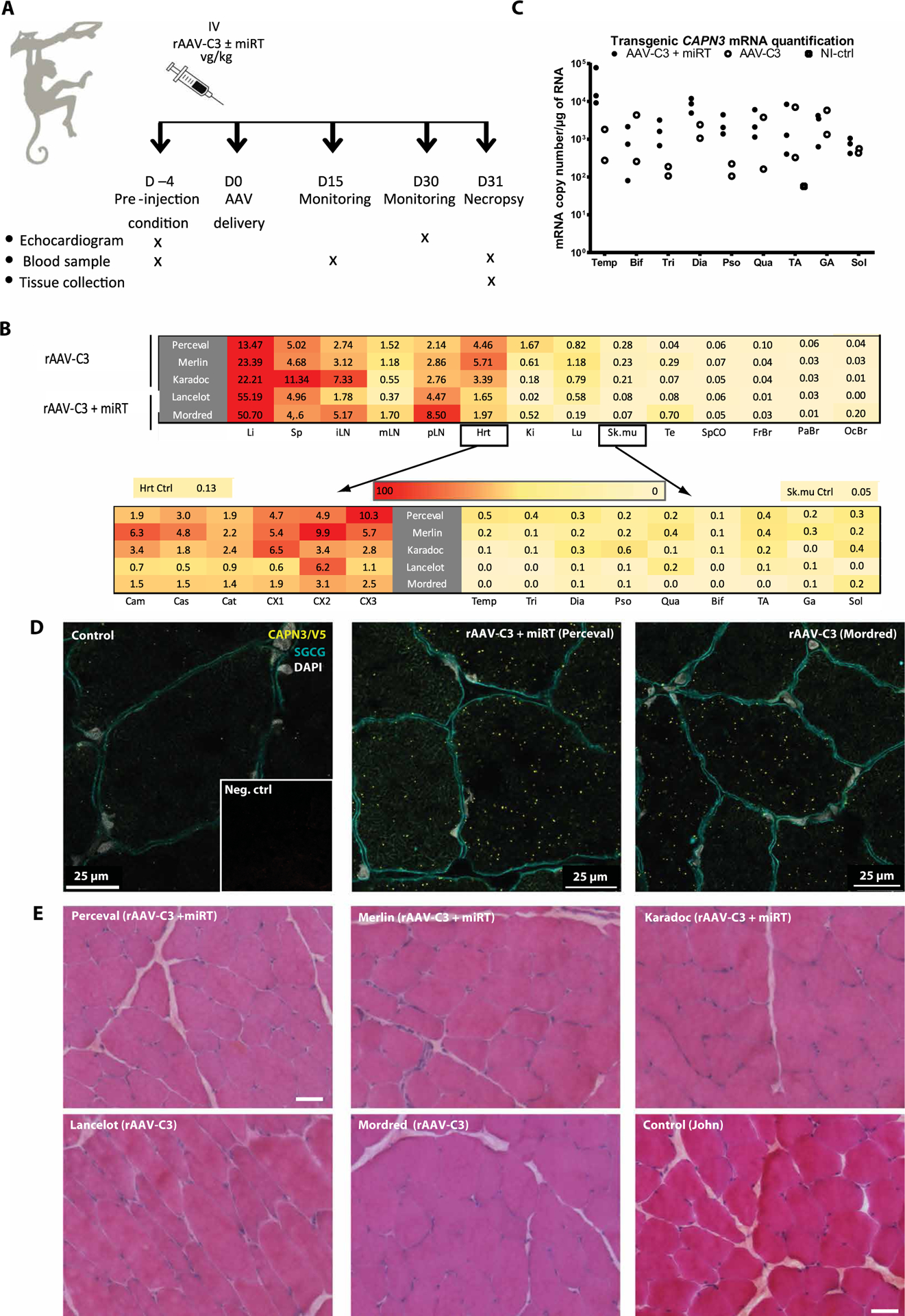
Fig. 3. NHP biodistribution study and transgene expression.

(A) Study design (n = 2 to 3). IV, intravenous injection. (B) Determination of vector copy number (VCN) per diploid genome in a range of tissues after systemic injection of rAAV-C3 or rAAV-C3+miRT sequences, represented by heat maps with a linear color table from red (high) to yellow (low). The values presented in the upper heat map for heart and skeletal muscle correspond to the mean of six different heart sections and the mean of all limb muscles, respectively, whose individual values are indicated in the lower heat maps. Li, liver; Sp, spleen; iLN, inguinal lymph nodes; mLN, mesenteric lymph nodes; pLN, popliteal lymph nodes; Hrt, heart; Ki, kidney; Lu, lung; Sk.mu, skeletal muscle; Te, testis; SpCO, spinal cord; FrBr, frontal brain section; PaBr, parietal brain section; OcBr, occipital brain section. (C) Expression of transgenic calpain 3 in a range of Macaca skeletal muscles (n = 2 to 3). Transgene expression was measured by RT-ddPCR and is presented in copies per microgram of RNA normalized to Rplp0. Temp, temporalis; Bif, biceps femoris; Tri, triceps brachii; Dia, diaphragm; Pso, psoas; Qua, quadriceps; GA, gastrocnemius; Sol, soleus. (D) Detection of transgenic calpain 3 using PLA. Red dots correspond to a specific amplification due to proximity ligation of two antibodies detecting epitopes present in our transgenic protein, specifically CAPN3 and V5 antibodies. Colabeling with γ-sarcoglycan (green) was performed to visualize muscle fibers, and 4′,6-diamidino-2-phenylindole (DAPI) is shown in blue. Inset: PLA negative control. Scale bars, 25 μm. (E) Sections of biceps femoris for all primates stained with HPS. Scale bars, 100 μm.
