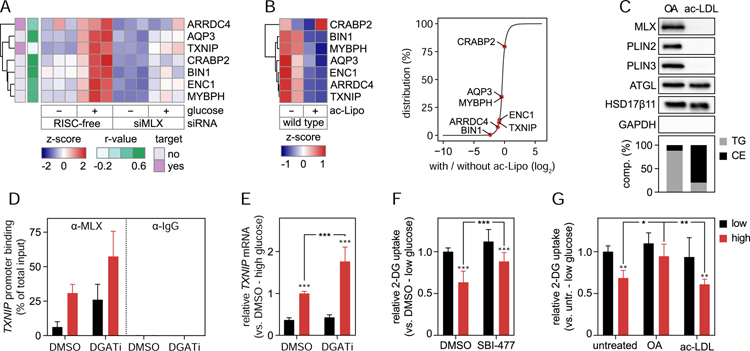
Figure 7. LDs modulate MLX:MLXIPL Gene Regulation in Human Macrophages.
(A–B) MLX controls a specific set of putative lipid storage response genes. (A) Genes depending on MLX for induction after glucose stimulus were identified by RNA sequencing. Control and MLX-depleted THP-1 macrophages were starved from glucose for 2 days and thereafter incubated in the presence or absence of glucose (25 mM) for 1 day. Known targets of MLX (TXNIP and ARRDC4) and genes displaying a similar LD phenotype as MLX in the RNAi screen (r>0.5) are highlighted. Results are based on three replicates per condition and were evaluated using DESeq2. (B) Heatmap and distribution chart of the effects of lipid storage induction by ac-Lipo on putative target gene expression. Counts from the RNA sequencing were scaled prior to clustering (left panel) and the relative position among all measured genes highlighted (right panel). Results are based on two replicates per condition.
(C) MLX binds specifically to TG-containing LDs. Based on fractionated THP-1 cells, protein and lipid composition of isolated LDs induced by incubations with OA (0.5 mM) or ac-LDL (25 µg/mL) for 2 days were determined using western blotting and TLC, respectively.
(D–E) LDs regulate MLX activity. TXNIP promoter binding of MLX and TXNIP mRNA levels were measured in THP-1 macrophages. Cells were starved from glucose for 1.5 days and thereafter incubated OA-containing media with or without glucose (10 mM in panel D and 25 mM in panel E) in the presence of DMSO or DGAT1/DGAT2 inhibitors for 12–16 hours. Results are based on one (D) or two (E) independent experiments, each containing three replicates and (E) were evaluated using two-way ANOVA followed by Tukey-Kramer test.
(F–G) Alterations in MLX:MLXIPL activity control 2-DG uptake. Uptake of [3H] 2-Deoxy-D-glucose in THP-1 macrophages was determined in cells (F) starved from glucose for 1 day and thereafter incubated with or without glucose in the presence of DMSO or 10 µM SBI-477 for 1 day and (G) incubated in varying concentrations of glucose in the presence or absence of OA (0.5 mM) or ac-LDL (25 µg/mL) for 2 days. Results are based on three (F) or two (G) independent experiments, each containing three replicates and were evaluated using two-way ANOVA followed by Tukey-Kramer test. Abbreviations: 2-DG, 2-deoxyglucose; ac-LDL, acetylated low-density lipoprotein; ac-Lipo, acetylated apolipoprotein B-containing lipoprotein; CE, cholesterol ester; comp., composition; OA, oleic acid; TG, triacylglycerol; untr., untreated.

