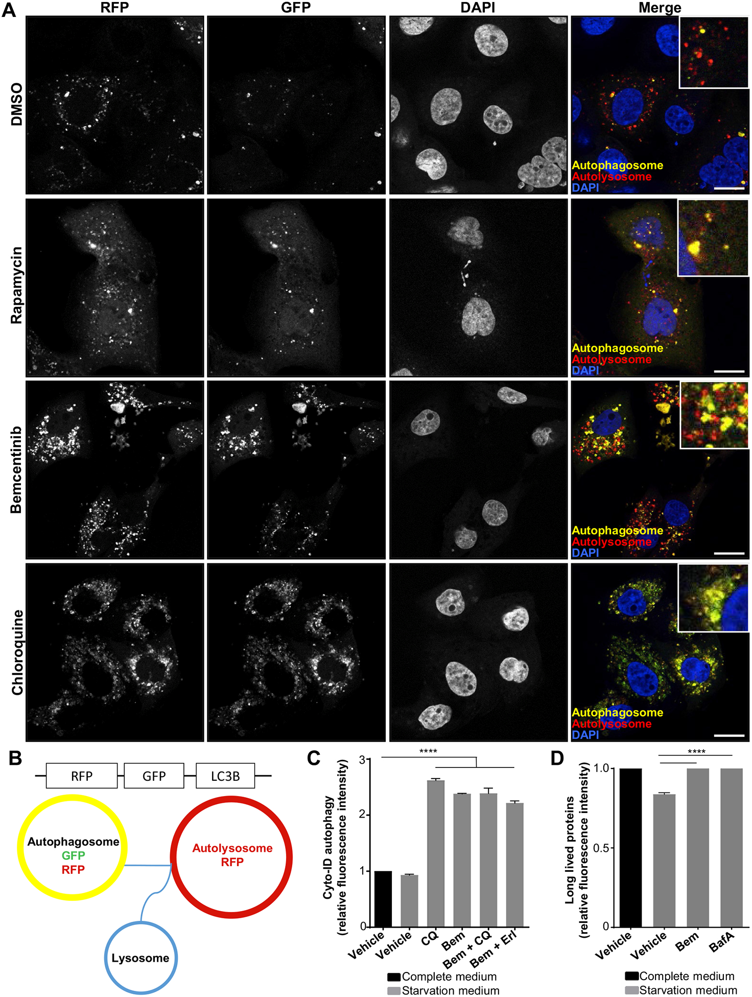
Figure 5.

The specific AXL inhibitor bemcentinib inhibits the high premortem autophagic flux of the erlotinib-resistant cells. (A) Dynamics of autophagosome formation and subsequent fusion with lysosomes to form autolysosomes was assessed by Premo Autophagy Tandem Sensor RFP-GFP-LC3B experiment. ER10 cells transfected with RFP-GFP-LC3B tandem sensor were treated as indicated for 24 hours with vehicle (DMSO), rapamycin (200 nM), bemcentinib (0.8 μM), or chloroquine (50 μM). Representative confocal images are shown. Scale bar: 10 μm. (B) Schematics for the assay described above. The LC3-associated autophagosomes seem yellow because of the simultaneous expression of both RFP and EGFP under neutral pH, (B) as on the fusion of the LC3-associated vesicles with the lysosome, the resultant autolysosomes express RFP only because of the loss of fluorescence from the acid–sensitive EGFP fluorochrome under acidic conditions. (C) Autophagic flux was assessed in ER3 cells by measuring the balance of autophagosome formation and clearance by pretreating the cells 16 hours (Vehicle [DMSO], 50 μM chloroquine [CQ], 1.2 μM bemcentinib, 10 μM erlotinib). Autophagy was induced through starvation in EBSS with 0.1% BSA for 3 hours (in the presence of treatment). Relative green fluorescence intensity from the Cyto-ID probe compared with the vehicle-treated cells in complete cell culture medium is given for a representative experiment ± SD (n = 3). Unpaired t test (****p < 0.0001). (D) Flow cytometry Click-IT AHA chemistry–based long-lived protein degradation assay of ER3 cells pretreated as indicated for 16 hours with bemcentinib 1.2 μM or Bafilomycin A 50 nM, before starvation in EBSS with 0.1% BSA for 3 hours (in the presence of treatment). The alkyne-AF488 fluorescence intensity ± SD (n = 3) was given for the starved samples relative to the vehicle-treated (DMSO) cells in complete RPMI-1640 cell culture medium. Unpaired t test (****p < 0.0001). BSA, bovine serum albumin; EBSS, Earle’s Balanced Salt Solution.
