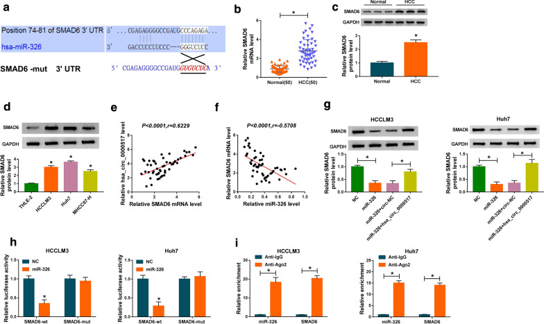
Fig. 5.
MiR-326 targeted SMAD6 in HCC cells. a The predicted binding sites between 3′ UTR of SMAD6 and miR-326 were predicted with targetscan database. b, c The levels of SMAD6 mRNA and protein in HCC tissues were detected via qRT-PCR or western blot analysis. d The level of SMAD6 protein in HCC cells was examined via western blot analysis. e, f The correlation between SMAD6 and hsa_circ_0000517 or miR-326 in HCC tissues was assessed with Pearson’s correlation analysis. g Effect of hsa_circ_0000517 overexpression on SMAD6 protein levels of miR-326-enhanced HCCLM3 and Huh7 cells was analyzed through western blot analysis. h Dual-luciferase reporter assay was executed to determine the luciferase intensity of the luciferase reporters containing SMAD6-wt or SMAD6-mut in HCCLM3 and Huh7 cells transfected with miR-326 or NC. i RIP assay was conducted to assess the miR-326 and SMAD6 enrichment in immunoprecipitates in HCCLM3 and Huh7 cells. *P < 0.05

