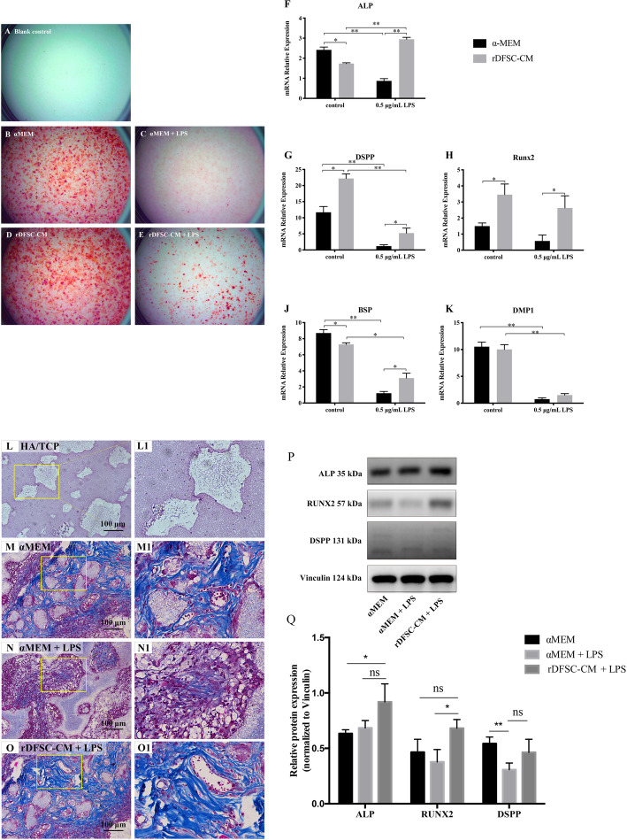
Fig. 6.
rDFSC-CM promoted the odontogenic differentiation and ectopic dentinogenesis of rDPCs.A–E Mineralized nodule formation of inflammatory rDPCs after odontogenic induction for 7 days. A Blank control group. B αMEM group. C αMEM + LPS group. D rDFSC-CM group. E rDFSC-CM + LPS group. Representative images of three independent experiments are shown. F–K Real-time PCR revealed that the treatment of inflammatory rDPCs with rDFSC-CM for 7 days upregulated the expression of the odontogenic genes ALP, DSPP, Runx2, BSP, and DMP1. β-Actin was used as an internal control. L–O Effects of rDFSC-CM on ectopic dentin collagen fibers of inflammatory rDPCs in vivo. The cells were loaded onto HA/TCP and then transplanted into the subcutaneous tissue of nude mice, where they remained for 8 weeks. The Masson staining results showed the production of collagen fibers (× 20). L, L1 Blank control group. M, M1 αMEM group. N, N1 αMEM + LPS group. O, O1 rDFSC-CM + LPS group. Representative images of three independent experiments are shown (scale bar 100 μm). P, Q As demonstrated by Western blot analysis, treatment with rDFSC-CM for 8 weeks significantly increased the expression of Runx2 in inflammatory rDPCs after in vivo. The levels of the ALP and DSPP proteins were also increased in the rDFSC-CM group, although the differences were not statistically significant. The molecular weights of the bands are indicated in kilodaltons. Vinculin was used as an internal control. The results are presented as the means ± SDs from at least three independent experiments. *P < 0.05, **P < 0.01; ns, no significant difference

