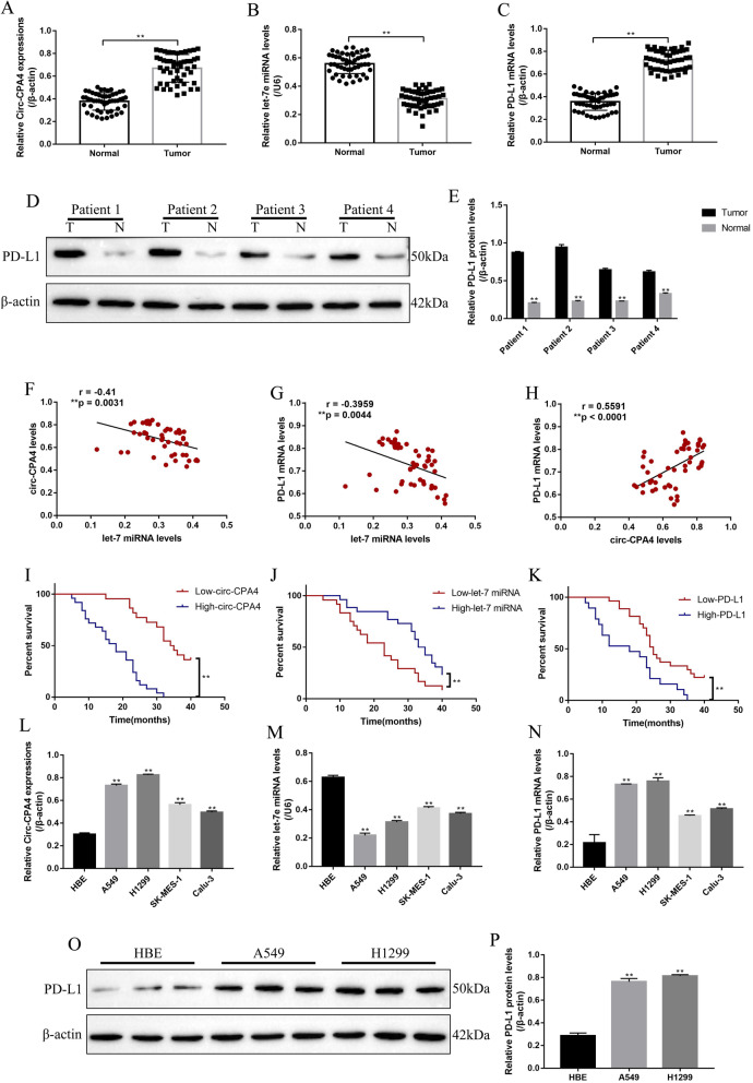
Fig. 1.
The expression status and correlation of circ-CPA4, let-7 miRNA and PD-L1 in NSCLC tissues and cell lines. The NSCLC tissues (N = 50) and their paired adjacent normal tissues (N = 50) were collected, and Real-Time qPCR was conducted to determine the expression levels of a circ-CPA4, b let-7 miRNA and c PD-L1 mRNA in NSCLC tissues and their paired adjacent normal tissues. d, e The clinical samples collected from four patients, and the expression levels of PD-L1 protein were measured by using Western Blot. (“T” represented “tumor tissues”, “N” represented “normal tissues”). Pearson correlation analysis was conducted to analyze the correlations of f circ-CPA4 and let-7 miRNA, g let-7 miRNA and PD-L1 mRNA and h circ-CPA4 and PD-L1 mRNA in NSCLC tissues. Kaplan-Meier survival analysis was performed to analyze the correlations of i circ-CPA4, j let-7 miRNA and (K) PD-L1 with NSCLC patients prognosis. Real-Time qPCR was used to measure the expressions of l circ-CPA4, m let-7 miRNA and n PD-L1 mRNA in the human NSCLC cell lines (A549, H1299, SK-MES-1 and Calu-3) and human normal bronchial epithelial cell line (HBE). o, p Western Blot was conducted to examine PD-L1 protein expressions in HBE, A549 and H1299 cells. Each experiment repeated at least 3 times. *P < 0.05, **P < 0.01, NS means no statistical significance

